Introduction
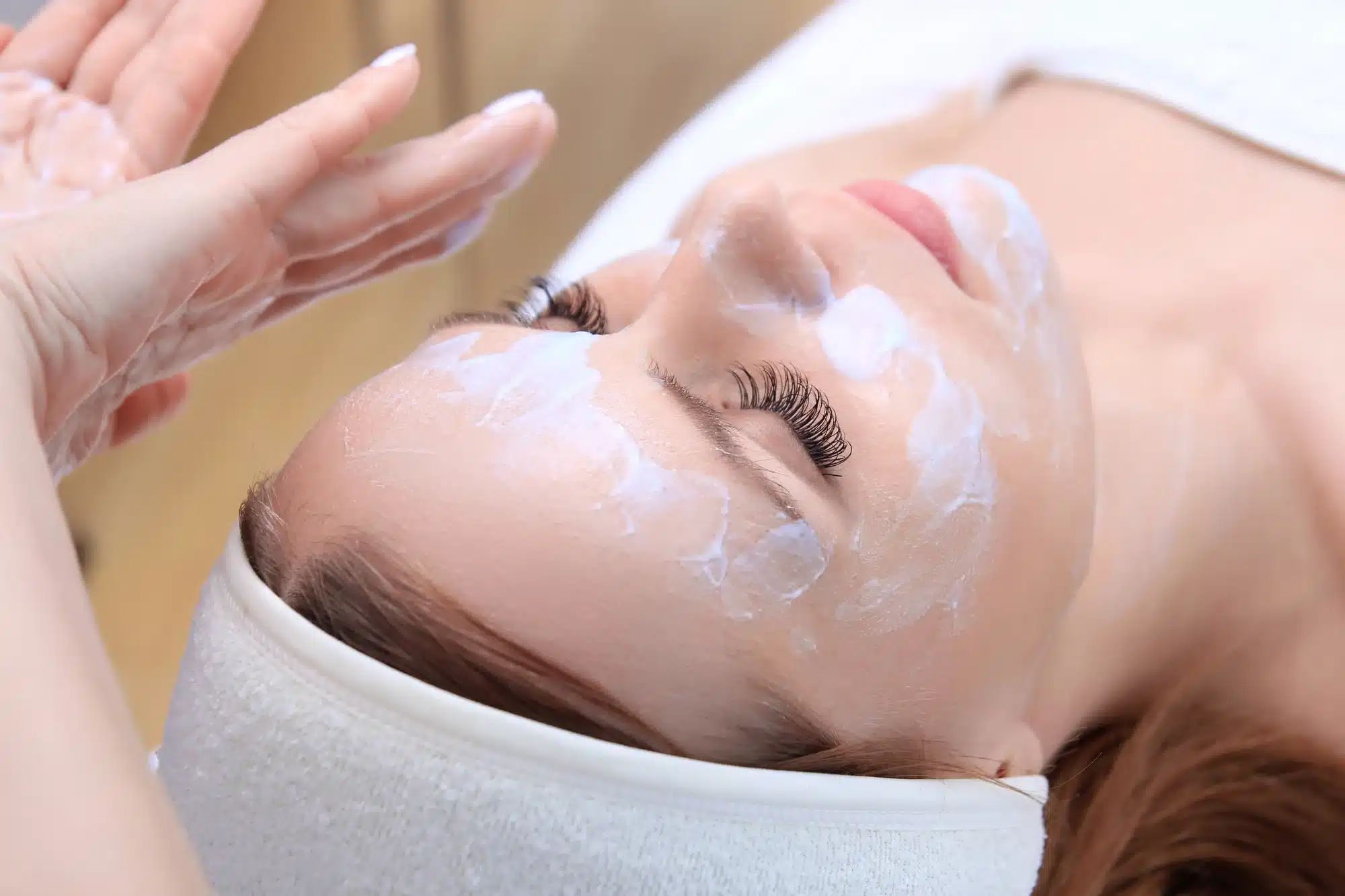
Chemical peeling, also known as chemexfoliation, is one of the oldest skin resurfacing treatments available. It involves the application of caustic agents to produce controlled, predictable destruction of the epidermis and variable portions of the dermis.1 The process is followed by regeneration and remodeling, with clinical improvement of texture and surface abnormality through subsequent re-epithelialization and second intention healing.
The advent of lasers put chemical peels on the back burner. Laser resurfacing is deemed favorable because it allows direct visualization of the depth of tissue ablation and tissue effects, while a peel event is an all-or-none approach.2 The results from both methods are operator-dependent, but lasers are more advantageous, particularly for moderate to severe photodamage. However, chemical peels have been making a comeback in the past few years, not only for their exfoliating properties and influence on collagen synthesis, but for their efficacy in treating common skin concerns.
For thousands of years, chemical peeling has evolved from utilizing natural products such as milk and honey to more caustic methods that treat a variety of indications including acne, photoaging, melasma, and scarring. With proper patient and product selection, patient counseling, adequate priming, and good pre-peel and post-peel care, one can prevent complications associated with chemical peeling and provide substantial effects comparable to lasers and radiofrequency.
Patient Analysis
As with any aesthetic procedure, proper patient selection is paramount. There are many reasons why patients seek cosmetic enhancement; thus, it is essential that the practitioner discuss what the procedure will achieve and what it will not. The most challenging aesthetic patient—and the one that requires the greatest degree of caution—is the person that presents with barely noticeable skin problems. These patients often have unrealistic expectations and are most likely to meticulously scrutinize results and be disappointed in what they may see as inadequate outcome. They may complain that they don’t see any skin improvements or that their faces “look the same.”
The physician must know each patient’s medical, social, and family history to identify possible contraindications to skin peeling. A detailed consent form should also be taken. Inquire about poor wound healing and the tendency to develop hypertrophic scars or post-inflammatory hyperpigmentation (PIH).3 Examine the patient in a well-lit room and with no make-up on. When treating acne scars, an overhead, movable light fixture is useful in examining the skin by shining indirect light to highlight certain scars. During physical examination, one should also eliminate the presence of skin disorders that have the tendency to spread to injured skin (psoriasis, lichen planus, and vitiligo). Furthermore, patients’ skin type (Fitzpatrick classification scale) and degree of photodamage (Glogau classification) should also be evaluated.4
To prevent complications, it is important to identify the patient at risk so that adverse effects can be anticipated and treated accordingly should they occur.5
- Patients with darker skin because they tend to develop PIH;
- Those with sensitive skin or a history of atopic dermatitis;
- Those with a history of photosensitivity or PIH;
- Those who work outdoors;
- Patients with dry skin and reddish hue;
- Patients with a history of poor wound healing, herpes infection, and/or keloids;
- Patients who were recently treated with isotretinoin;
- Patients with unrealistic expectations;
- Uncooperative patients or those who are psychologically disturbed
Indications for Chemical Peels
As basic understanding of the peeling process has advanced and a greater number of agents have been made available, skin peeling has become not only a method of reversing photodamage but also an essential option in treating a variety of dermatologic conditions. The most important aspect of skin resurfacing is deciding the proper level of peeling, which is not too deeper nor too superficial. The key to selecting the proper level of resurfacing is having a thorough understanding of skin anatomy and the depth of the patientâs skin concern. Peels are traditionally classified based on their depth of penetration: very superficial, superficial, medium, and deep. Some consider this classification obsolete since the depth of peeling is dependent on several factors such as the concentration and pH of the solution, the number of layers applied, and the contact time. Philippe Deprez described the seven possible depths of peeling:6
- Exfoliation (very secure zone)
The first depth is the most superficial and consists of simple peeling of dead cells in the stratum corneum, revealing superficial keratinocytes. These are living cells that contain more water than stratum corneum cells. Alpha hydroxy acid (AHA) solutions are primarily used for this depth.
- Intraepidermal peel (very secure zone)
These peel solutions penetrate the epidermis, removing more cells, but do not affect any part of the dermis or even the basal layer. Depth 2 peels give better results than depth 1 peels and are useful for treating many keratinization problems and epidermal melasma. Common agents for intraepidermal peels include AHA, trichloroacetic acid (TCA), alpha ketoacids, resorcine, and salicylic acid.
- Basal dermis peel (still secure)
In this peeling level, the stratum corneum cells are removed completely and keratinocytes in the basal layer are largely damaged. Still, the epidermis is not completely destroyed as many keratinocytes reside in the deep epidermal papillae, which are less prone to UV damage; hence, skin can easily regenerate. Basal dermis peels treat aging skin, acne, fine lines, melasma, and keratoses. TCA is the best agent for this depth.
- Grenz zone peel (still secure)
This peel presents a low level of risk, is not very painful for the patient, and provides good results. TCAs are preferred over AHAs because the latter may cause irregular penetration. Acids penetrate the superficial layers of the papillary dermis, destroying stratum corneum and a large portion of keratinocytes, eliminating abnormal epidermal cells; thus, this type of peel is an effective treatment for melasma, lentigines, and keratoses.
- Papillary dermis peel (quite secure)
This peel is advantageous in treating many skin conditions, such as solar keratoses, melasma, lentigines, and fine lines. However, it borders between secure and insecure depths. Salicylic acid, AHA, resorcine, and alpha ketoacid are not recommended for this depth. TCA is still the best choice for a safe and effective papillary dermis peeling.
- Reticular dermis peel (dangerous)
This peel is regarded as the Holy Grail of peeling as it can treat nearly all pigment defects and wrinkles. However, it is not the best peel for patients with thick and oily skin because this type of skin resists the action of acids. TCAs and phenol are mainly used for this depth.
Skin Considerations and Contraindications
Thanks to newly developed carrier solutions, caustic agents and secondary ingredients are now transported across the stratum corneum (protected from degradation) into the deep layers of the skin to target skin concerns. It is recommended to use the same ingredients for the peel and creams used in pre-, post, and during peel treatment, so that the peel will boost the activity of the creams.7-11
Chemical peeling poses an advantage for most skin types over other resurfacing techniques if proper skin conditioning and proper procedure depth are respected. However, patients with darker skin are at risk for permanent hypopigmentation using peels that penetrate the depth of the reticular dermis. This skin type is also at risk for post-inflammatory hyperpigmentation with any peel level. The length of pre-peel skin conditioning should be extended to three months to minimize the risk, resuming immediately upon re-epithelialization of the skin.
Fitzpatrick Phototypes
Fitzpatrick Phototypes classify skin type based on its response to ultraviolet exposure. Patients are categorized from I – VI according to the progression of skin darkening and as their ability to tan rather than burn increases. While it helps clinicians treat patients with phototherapy, this classification is limited because it does not address the degree of photodamage present or aid in choosing the correct peel depth.
Fitzpatrick Phototype Chart
| Blonde or red hair, freckles, blue or gray eyes, very pale skin, burns very easily and never tans | High risk for vascular damage and skin cancer. Often develop obvious erythema on any unprotected exposure to the sun’s rays. May scar if slow to heal. |
| Sandy or red hair, fair skin with some freckles, burns easily and tans with difficulty | High potential for vascular damage and high risk for skin cancer. May scar if slow to heal. May develop pigmentation with trauma. |
| Brown or sandy hair, fair skin, hazel, green, or blue eyes, slow to burn, develops a deep even tan | High potential for scarring and pigmented skin conditions. Moderate risk for vascular damage and skin cancer. |
| Dark brown or black hair, green, hazel, or brown eyes, olive complexion, tans easily and rarely burns | High risk for scarring and pigmentation caused by trauma, heat, and chemicals. Moderate risk for visible vascular damage. |
| Dark or black hair, brown or dark brown eyes, very dark complexion, may never burn | High risk for keloid scarring and pigmentation caused by trauma, heat, and chemicals. Moderate risk for visible vascular damage. Lower risk for solar pigmented skin conditions. |
| Black hair, dark brown or black eyes, may never burn | Very high risk for keloid scarring and pigmentation caused by trauma, heat, and chemicals. Moderate risk for visible vascular damage. Lower risk for solar pigmented skin conditions. |
Glogau’s Classification
Glogau’s classification objectively quantifies the amount of photodamage present using four age-range levels but does not help in determining the depth of resurfacing needed and best peeling modality. Glogau is an easy-to-use clinical scale that categorizes wrinkle severity and provides a useful measure of treatment effectivity.12 Both the Fitzpatrick and Glogau scale were designed prior to the introduction of non-ablative techniques and fall short in addressing thicker skin or darker skin types.
| Classification | Typical Age | Characteristics |
| Mild (No wrinkles) | 28-35 | Small or no wrinkles, early photo-aging, without keratosis |
| Moderate (Wrinkles in motion) | 35-50 | Small wrinkles, early to moderate photo-aging, appearance of lines when the face is in motion, early brown spots visible, sallow complexion, with actinic keratosis |
| Advanced (Wrinkles at rest) | 50-65 | Deep wrinkles, advanced photoaging or discoloration, prominent capillaries and brown pigmentation, visible keratosis, wrinkles now present at rest |
| Severe (Only wrinkles) | 60 above | Dynamic and gravitational wrinkles, severe photoaging, yellow-gray complexion, actinic keratosis |
Skin Ethnicity
Skin color alone is not enough to guide the peeling depth. Fanous stated that facial features were more important than skin color in determining PIH risk in patients with a mixed-race background.13 Arab skin type, for example, is more prone to PIH while Celtic skin has high erythema sensitivity. If a patient has a Celtic mother and an Arab father, PIH risk is based on which parent the patient looks most like, regardless of their skin color.
Other Considerations
- Patients with a history of immunosuppression, autoimmune disease, exposure to radiation, and collagen vascular disease as well as isotretinoin could compromise the healing capacity of the skin. Before undergoing chemical peeling, it is recommended to wait 12 months from the end of isotretinoin therapy.
- If a patient has sensitive skin, perform a patch test in a small inconspicuous location before embarking on a full-face peel.
- If a patient has poor skin condition (pigmentation problems, low hydration, damaged blood vessels, etc.), reschedule the peel for another time and use creams or serums instead to boost skin structure, repair damage, and to appropriately prepare the skin
- It is important for the skin to be properly hydrated prior to treatment, as peels need water to move through the skin. Poorly hydrated skin is more likely to react to the hydrogen ions of the peel and may experience localized damage to the surface, leading to an increased risk of adverse reactions.
Peel Exposure Time
Exposure time is the time during which the peeling agent is left to act on the skin before being neutralized, stopping its effect. Due to the ability to transport resurfacing agents into the skin using specialized carrier technology, surface trauma that is normally seen with traditional peels is greatly reduced. Although contact time should be set prior to application, the visible symptoms of the peel and the patient’s response should still be considered. For example, when using AHA, contact time should end as soon as erythema begins. However, AHAs in aqueous solution do not penetrate the epidermis evenly and erythema may appear in spots or patches. The safety limits are not very flexible when using AHA peels in aqueous solution; thus, the physician must stay beside the patient and carefully observe the treated area, as this peel can go from superficial to deep very quickly.
Conclusion
Chemical peeling has withstood the test of time and continues to play an important role in skin resurfacing. The allure of chemexfoliation lies in its ability to treat patients of varying skin types, as well as for allowing the procedure to be tailored to different depths on different areas of the face. As with any cosmetic procedure, optimal results depend on proper procedure selection, patient evaluation, and the utilization of appropriate skin care regimens, including the use of products such as Belotero.
References
- Brody, H.J. (1997). Histology and classification in: Brody HG ed Chemical Peeling and Resurfacing, Mosby Yearbook, pp. 1-6
- Kauvar, A., & Dover, S. (2001). Facial rejuvenation: Laser resurfacing or chemical peel: choose your weapon, Dermatol Surg, 27, 2.
- Obagi, S., & Niamtu, J. (2016). Cosmetic Facial Surgery: Chemical Peel, Elsevier Health Sciences.
- Lee, J., Daniels, M., & Roth, M. (2016). Mesotherapy, microneedling, and chemical peels. Clin Plastic Surg, 43.
- Khunger, N. (2009). Complications. In: Step by step chemical peels. New Delhi: Jaypee Medical Publishers, pp. 280-97
- Deprez, P. (2017). Textbook of Chemical Peels, CRC Press.
- Shuller, P., & Mayr-Kanhaauser (2003). Enerpeel 50% Pyruvic Acid Comparison with 70% Glycolic Acid – UnivGraz, Austria.
- Bonina (2003). Enerpeel PA efficacy and safety evaluation versus 50% pyruvic acid. Univ Catania, Italy.
- Beradesca, M., & Gallicano, S. (2004). Clinical evaluation of Enerpeel PA, Derm Institute, 3.
- Berardesca, E., Cameli, N., Primavera, G., & Carrera, M. (2006). Evaluation of Skin Improvement after Treatment with a New 50% Pyruvic Acid Peel, Dermatol Surg, 32, 4.
- Raone, B., & Patrizi, A. (2017). Salicylic acid peel incorporating Triethyl citrate and Ethyl linoleate in the treatment of acne: a new therapeutic approach, Internal Medicine, Aging and Nephrologic Diseases Department, Division of Dermatology, Sant’Orsola-Malpighi Hospital, University of Bologna, Bologna, Italy.
- Glogau, R.G. (1996). Aesthetic and anatomic analysis of the aging skin, Med. Surg, 15.
- Fanous, N. (2002). A new patient classification for laser resurfacing and peels: Predicting responses, risks and results. Aest Plast Surg, 26.
Related Articles
Joanna Carr
Depo Provera Effectiveness – What Are the Chances of Still Getting Pregnant?
Expert opinions and clinical studies support the reliability of Depo Provera as a contraceptive method. Read more on Doctor Medica.
Joanna Carr
Lanluma Side Effects – Short Term, Long Term, Mild, Serious
Understand Lanluma’s potential side effects—from mild, short-term reactions to rare, serious complications. Learn what to expect and how to manage pos...
Joanna Carr
Diamond Injection Technique for Neck and Décolleté Rejuvenation
Discover the Diamond Technique for neck and décolleté rejuvenation! Learn how this innovative approach enhances collagen and improves skin texture.


