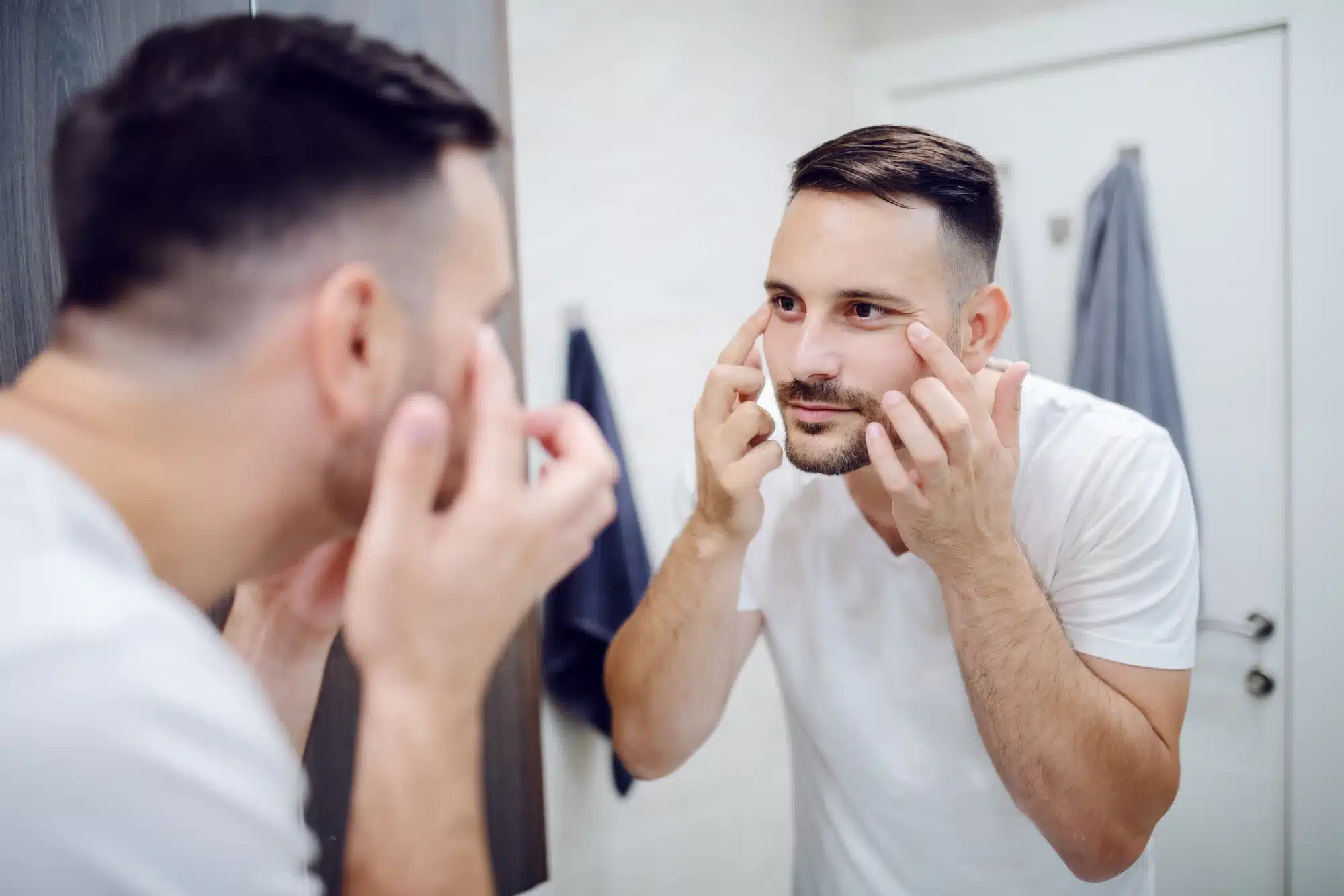
What is a tear trough deformity?
The tear trough area refers to the skin that radiates outwards and downwards from the inner corner of the eye. A youthful tear trough should have a relatively smooth transition between the preseptal and orbital parts of the orbicularis oculi (OO) muscle; it should also continue to the upper malar region without any obvious transition point. This region is made of extremely thin and delicate skin that is bound to display various signs of aging due to its fragile nature. Some of the commonly reported tear trough deformities are dark circles and deep lines. Patients with prominent tear trough deformities can have a sunken appearance in the lower eyelid region, which will inadvertently cast a dark shadow that cannot be masked using cosmetic products, such as concealer. Aesthetic deformities of the tear trough usually happen due to a combination of several factors, including a loss of volume associated with aging, laxity and changes in skin thickness, hyperpigmentation, noticeable pooling of venous superficial vessels, and patients’ ethnicities. If left uncorrected, tear trough imperfections can make a person look exhausted, despite adequate resting, and significantly older than their actual age.
What are the different classes of tear trough deformity?
For easy identification and treatment, tear trough deformities are classified based on their severity. The following classification is established by Hirmand and ranges from class 1 (mild) to class 3 (severe):
- Class 1 (Mild): Patients with a loss of volume that is limited medically to the tear trough and has mild flattening that extends to the central cheek are categorized into this class.
- Class 2 (Moderate): Patients are diagnosed with moderate tear trough deformities when they display loss of volume in the lateral and medial orbital areas. They may also have a moderate deficiency of volume in the medial cheek and a flattened central upper cheek.
- Class 3 (Severe): Patients who experience full circumferential depression along the orbital rim that extends from the medial to lateral areas are diagnosed with severe tear trough deformities.
Estheticians are encouraged to discuss the severity of deformities using picture examples for clearer understanding with their patients. Picture examples can also be used to explain the possible and realistic outcomes of a tear trough procedure. It is imperative that patients be made to understand that the tear trough with severe cases of deformities cannot be fully restored to the way it appeared when the patient was younger without the need for multiple treatment sessions or surgery.
What are the treatment options for a tear trough deformity?
While most patients use topical cosmeceuticals to treat their tear trough issues, a number of aesthetic procedures have been developed to effectively banish these imperfections at a much faster rate. Some suitable cosmetic procedures include microneedling, platelet rich plasma (PRP), and injecting the tear trough region with dermal fillers. Estheticians are encouraged to use hyaluronic acid-based implants like Teosyal Redensity II (RD2), as it is specifically developed to treat aging signs in the under-eye area. Once it is injected correctly, the hyaluronic acid filler has a lower risk of migration and causing lumps than is usually seen with other brands. This filler is also renowned for not causing a significant amount of swelling in the treated area. Teosyal Redensity II has a distinctive viscoelastic property thanks to the presence of both cross-linked and non-crosslinked hyaluronic acid chains. The viscoelastic feature of the filler makes it easy to integrate uniformly into the tissues. For most patients, a single 1ml preloaded syringe is enough to correct both eyes in just one sitting. The injection techniques may vary depending on the patient’s age and skin health. Younger patients with an even skin tone and very little laxity can be treated via the serial point injection technique. This treatment approach involves administering the filler using a 30G needle in multiple and evenly spaced injections. Older patients aged above 50 are best treated using a 27G or 25G cannula, so as to reduce the risk of bruising. The risk of intravascular injury is quite rare in this tear trough region as long as the physicians avoid the angular artery, which is the terminal branch of the facial artery. This angular artery travels along the nose to the inner canthus to supply blood to the upper and lower eyelids and the nose. That being said, anatomical variations of vessels can still occur. Therefore, practitioners must have an in-depth anatomical knowledge of the tear trough region. Prior to starting the treatment session, patients must be informed on all possible outcomes; similarly, rare adverse health reactions, like ocular blindness, must be disclosed to the patients before the treatment.
How are tear trough deformities treated using dermal fillers?
Just like any other aesthetic procedures, dermal filler injections can only be done once patients have been counselled about the possible outcomes, warnings, and precautions. Injectors must also obtain a patient’s informed consent before starting the treatment. Commence the session by cleaning and disinfecting the tear trough region. Patients should be seated at a 45° angle; lying flat will only mask the deformities and make the targeted injections more difficult. They are also advised to keep their eyes opened whilst looking upwards. This stops them from squeezing their eyes tightly and raising their cheeks during the injecting process, which can also obscure the imperfections. Injectors are advised to treat the deeper tear trough deformity first. They should insert the needle, pointed bevel up and away from the eye, at the most lateral part of the tear trough region and perpendicular to the skin. The filler must be injected into this delicate area slowly and with very minimal movements. Inject about 0.05ml to 0.1ml of hyaluronic acid gel in the tissues above the bone but below the orbicularis oris (OO) muscles. Though superficial injection of the filler into the OO muscle does result in instant plumping of the area, this method can actually cause filler migration, as the OO muscle contracts and displaces the gel medially. Remove the needle and apply gentle pressure immediately before continuing injecting the region in a medial direction. Once the area has been treated adequately, gently massage it to encourage smooth integration of the gel with the neighboring tissues. This serial point method is ideal for treating mild to moderate imperfections.
If estheticians are using a cannula to administer the filler gel, then the entry point should be just lateral and below the area where the tear trough ends. It is imperative to confirm that the cannula is of sufficient length so that it can reach the canthus region easily. The appropriate depth of the cannula insertion can be easily determined by checking for tenting caused by the cannula shaft once it is injected into the skin. An obvious skin tent simply means that the depth is too superficial. In this case, withdraw the cannula and reinsert it with a deeper angle below the OO muscle until no tenting is observed. Physicians should slowly inject small boluses of filler in the tear trough line with only small gaps between each injection. Doing so will allow the filler to expand during hydration and reduce the risk of overfilling. Once the area has been corrected, discard any leftover gel and used medical supplies. Patients are advised to keep the area clean and away from sunlight. Patients must also avoid alcohol and stay upright for four hours after the injection. They must avoid strenuous physical activities for the first 48 to 72 hours to reduce the risk of accidental bruising. Post-injection swelling can be effectively curbed using antihistamines. Practitioners must cease the treatment session straightaway if there are any concerns from the patients.
What are the results of tear trough augmentation using dermal filler?
After completion of the treatment procedure, patients should return for a follow up session about four weeks from the date of the initial session. By then, the localized inflammatory reactions, such as bruising and swelling, will have dissipated. A majority of patients will notice a tremendous improvement to their dark lines after the initial appointment; the periorbital skin area will also be brighter and more radiant. Leading up to follow up sessions, patients have the option to receive more dermal filler injections, depending on what their skin condition is and preferences are. Most hyaluronic acid-based implants should last for about six to nine months in the tear trough region before they succumb to degradation caused by the surrounding tissues. Fillers that are injected deep into the orbicularis muscle can actually last much longer with a duration of about 12 to 15 months since the hyaluronic acid degrades at a much slower rate in that region. However, this involves an advanced injection technique, and estheticians are advised to attend workshops on the matter that are organized by the product manufacturers. This is to ensure that the health-care specialists are equipped with extensive anatomical knowledge and are competent in both the injection technique and how to manage any potential complications.
Related Articles
Joanna Carr
SoftFil Precision – A Guide to This Advanced Dermal Filler Technique
Explore Softfil Precision—a comprehensive guide to this advanced dermal filler technique, designed for precise and natural-looking results in facial r...
Joanna Carr
Profhilo Lumps – What Are They and How to Deal With Them
Profhilo is an injectable hyaluronic acid treatment that stimulates collagen and elastin production, producing firmer and smoother skin.
Joanna Carr
Filorga: Quenching Thirsty Skin with Hyaluronic Acid
Revitalize your skin with Filorga's hyaluronic acid treatments for ultimate hydration. Explore Doctor Medica's range of products today!


