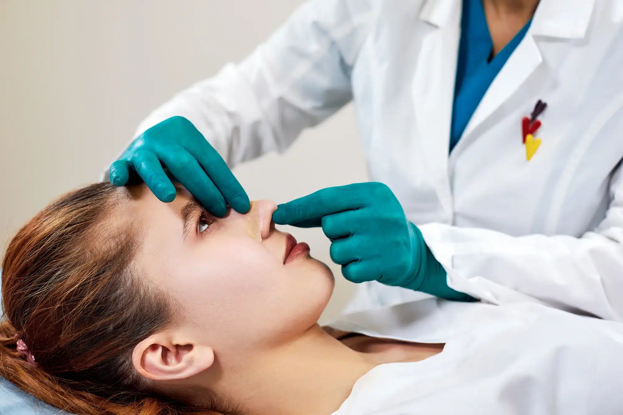
Nonsurgical Rhinoplasty
Rhinoplasty is increasingly in demand as more patients find themselves aware of the ideal nose appearance through the media. Even relatively small issues warrant a visit to an expert practitioner for the necessary corrections. The appeal of existing techniques that use implants and autologous cartilages are hampered by their long recovery times, high cost, implant-related complications, and the psychological hurdle(s) that patients need to overcome. The steep learning curve for rhinoplasty has also hindered many patients from reliably finding a suitable practitioner. This has led to an increase in popularity of nonsurgical rhinoplasty (NSR). NSR is the umbrella term that describes rhinoplasty procedures that do not require significant downtime and do not have the major risk for complications associated with surgery. One of the examples of NSR is the use of fillers, particularly hyaluronic acid. Fillers have become a method of choice for volume augmentation due to their safety profile, ease of use, and low cost. This article is the first of three parts that will cover nonsurgical rhinoplasty.
Treatment considerations
- High-risk areas have a higher risk for complications. Have hyaluronidase prepared and ready to be administered in the case of intravascular injections. Also, be familiar with its doses and dilution factors.
- Indications to abandon NSR and opt for surgical correction can be found when the tip does not move and is hard when the nasolabial junction is pinched.
- When you determine that patients require implants, use only small aliquots of hyaluronic acid (0.1ml to 0.2ml) to blend in the surrounding irregularities.
- Understand that compromising the blood supply (via inadvertent intravascular injection) and arteries that were fixed by scar tissue substantially increases the risk of vascular compromise, which can result in skin and cartilage necrosis. Exercise great caution when dealing with these areas of concern. Reconsider treating less pliable tissue and tissue that is more difficult to inject during the procedure.
Treatment techniques
The key of successful NSR is knowing its limitations. Know that some nose deformities can be treated with NSR, while others can only be treated with surgical techniques. Ideal deformities for using the NSR technique are ones that confer a minimal chance for complications. Examples include saddle deformity, mild dorsal hump, flat nose, and short nose. Less than ideal deformities, on the other hand, are high-risk areas that should only be approached by practitioners with the required technical know-how and experience of handling complications that are more likely to occur. High-risk areas include the alar recess, micronose, nasal tip, and glabella.
Both a needle or cannula are widely acceptable methods to administer dermal fillers for NSR.
Approaches to NSR
1. Spreader grafts
Functional improvements can also be obtained when the dermal fillers are injected to widen the nasal valve to increase the passage of air. In the past, this was performed as a surgical procedure by harvesting and inserting cartilage grafts to lift the upper lateral cartilages off the septum. Nowadays, hyaluronic acid and hydroxyapatite can achieve it without the technicalities involved in invasive surgery, though both of these are only temporary solutions to a permanent problem.
2. Nasal tip
First, introduce the dermal filler material to the intradermal area for a bifid tip. This can also be done to widen the nasal lobule if necessary. Note that there is no foundation to which the filler material can rest on if it is injected into the nasal tip. The augmentation will not last long, and it can create a predisposition to vascular compromise. Studies on cadaveric specimens have found that the midline longitudinal columellar artery is present in 31.1% of the performed dissections. The high-risk, low-reward ratio places this area as less than ideal as the site of injection. A good alternative injection site is between the anterior nasal spine at the base of the columella and the footplates of the lower lateral cartilages. A range of 0.5–1ml of filler material may be adequate for this area. If you find that the tip is hard, thick, and difficult to move when the nasolabial junction is pinched, identify these patients as requiring a surgical procedure. Inject about 0.5ml into the columella to provide support for the tip of the nose.
3. Cantilever nose lift
Begin by introducing 2% lidocaine at the site of correction. After that, insert the needle past the sellion into the nasal tip for dorsal augmentation. For nasal tip elevation, go posteriorly into the nasal spine with a 22G 55mm blunt cannula.
The dermal material should be inserted into the supraperiosteal plane via a deep subcutaneous layer injection. The cannula must be inserted into the nasion at the dorsum and past the sellion. To check the position of the cannula tip, you can pinch the dorsum of the nose. It is also useful later when you need to mold the product to avoid lateral spreading. Gently deposit the material using the retrograde injection technique because this will ensure that the product stays in the appropriate plane.
If there is ever a need for the dorsum to be wider, do not add volume in the single tunnel as created by the cannula. Just retract the cannula and form a new lateral and inferior tunnel to the original tunnel. Repeat this for the other side.
It is important to note ethnic differences in anatomy. The sellion can be found higher in the Caucasian face than other ethnicities. Dorsum injections can move the sellion up to 5mm closer in line with the medial brow. Assess the rhinion before deciding if the material should be injected at this area. In the case of a small dorsal hump, proceed to inject at the cephalic and caudal part of the hump.
To complete the treatment, simply augment the medial brow using a 30G needle. Take great caution as to not inject into the blood vessels. Aspirate the syringe before injection and repeat the retrograde technique. Contour the glabella so that it is consistent with the upper nasal dorsum. Injecting the medial brow to the radix ensures a smooth arch and also softens frown lines at the glabellar region.
4. Columella recession
Starting at the nasal tip, place the cannula posteriorly down the columella in the midline and perform the retrograde injection technique. Use multiple tunnels if necessary, but only up to a maximum of 0.5ml. Next, use a 30G needle to enter the nasal spine and aim for a nasolabial angle between 110° to 120°. This is commonly done alongside columella correction. Up to a total of 1ml can be used in these areas.
NSR complications
Cases of intravascular injection of the cosmetic filler material leading to the embolism of the the eyes and brain and skin necrosis have been well-documented. The latter has two distinctive factors: intravascular, where the hyaluronic acid fillers cause direct obstruction and chemical damage to the endothelial lining, and extravascular, where external venous compression from excessive volume injection, edema, or local inflammatory response impedes blood flow to the area. Most cases of embolism occur as a result of injection directly into the dorsal nasal artery or the lateral nasal artery. The dorsal nasal artery lies at the dorsum of the nose at about 3 mm away from the midline. If the needle is inserted into it and dermal filler material injected, it will travel down through its anastomotic connections, which are the ophthalmic, infratrochlear, and angular arteries. The area of necrosis will reflect these areas of anastomosis, and when it enters the ophthalmic circulation, certain clinical effects, such as sudden blind spots or visual field deficit, will occur.
Signs of intraarterial injection
Recognizing the signs of intraarterial injection is vital in salvaging the procedure. Patients will feel excruciating pain, and sometimes, they will complain of a sensation of something spreading out from the injection site. The ischemic area will develop edema within several hours. The skin will then start to blanch, or blotchy red patches may begin to form due to venous congestion as a rebound phenomenon. Capillary refill time will be prolonged and slow due to decreased arterial blood supply. After one day, ulcerative lesions will appear with the erythema, resulting in tissue desquamation. It will worsen for some time thereafter, with definitive findings of dermal necrosis (i.e. eschar formation) eventually occurring. The skin can recover through the wound healing process.
Other complications include:
- Tyndall effect: If you place the dermal filler too superficially, a bluish discoloration will result when the skin is illuminated. This is due to light scattering through the colloid solution. It can be corrected with hyaluronidase.
- Infection: Maintain aseptic techniques and standards throughout the procedure through adequate skin preparation and disinfectant agents. Broad-spectrum antibiotics, such as ofloxacin, can be utilized if the patient develops an infection.
- Lumps and nodules: These can be either granulomas or inflammatory and/or non-inflammatory nodules. Granulomas are an immune-mediated reaction in response to the injected foreign material; it is therefore an accumulation of immune cells that are trying to eliminate the dermal filler. It can be treated with corticosteroid injections or surgical removal. Inflammatory nodules can appear in days or in years, whereas non-inflammatory nodules appear immediately after injection and is most commonly caused by improper filler placement.
Management of complications
Most complications from hyaluronic acid dermal fillers can be reversed with hyaluronidase. It is imperative that you have the expertise to use it, especially in the case of emergencies. As mentioned earlier, the severe consequences associated with an intravascular injection can be best treated with an immediate injection of hyaluronidase into the area. Hyaluronidase should be directed into the affected vessel or the surrounding tissues, as some have reported that it may diffuse through the tunica intima. Different hyaluronic acid dermal filler formulations require different doses of hyaluronidase to dissolve. In addition, use 2% nitroglycerin pastes, hot packs, and soft massages in an attempt at vasodilation of the affected area. Despite the potential for reversibility, cosmetic fillers should always be applied via sound technique and comprehensive knowledge of the anatomy of the nasal structure to prevent complications in the first place.
Conclusion
The rate of advancements in this part of the cosmetic facial surgery field will most likely continue to occur for the foreseeable future. As more and more options are available for both practitioners and patients alike, the industry continues to progress in the availability of minimally invasive treatments that are safer and longer-lasting than surgical procedures. However, treatment with dermal fillers carries a litany of extreme complications, which gives focus to a central tenet: all specialists licensed to inject these products must become experts of human anatomy and proper application technique. With the patient’s life possibly at risk, there is always a need for practitioners to remain astute throughout the procedure to improve outcome.
Related Articles
Joanna Carr
Revanesse Versa Before And After With Photos
Revanesse Versa is a hyaluronic acid-based dermal filler known for delivering smooth, natural-looking results in various facial areas, including wrink...
Joanna Carr
Stylage vs Juvederm: Similarities and Differences Reviewed
Have an interest in learning about Stylage vs Juvederm And Their Similarities & Differences Reviewed? Browse Doctor Medica's extensive archive of blog...
Joanna Carr
Treating Permanent Dermal Filler Complications
Have an interest in learning about Treating Permanent Dermal Filler Complications? Browse Doctor Medica's extensive archive of blog postings.


